
Meta Description: Discover everything about PFN nail surgery for hip fractures—procedure details, recovery timeline, PFN vs PFNA comparison, and clinical outcomes in this comprehensive orthopedic guide.
Introduction
Intertrochanteric femur fractures represent one of the most common orthopedic injuries in the elderly population, accounting for approximately 50% of all hip fractures. With global hip fracture numbers projected to reach 4.5 million by 2050, the demand for effective surgical fixation continues to rise.
The Proximal Femoral Nail (PFN) has emerged as a cornerstone in the treatment of these challenging fractures. Developed by the AO/ASIF in 1996, this intramedullary device was specifically engineered to address the limitations of earlier extramedullary fixation methods. Today, PFN nail surgery represents a minimally invasive, biomechanically stable solution that enables early mobilization and improved patient outcomes.
This comprehensive guide explores everything you need to know about PFN nail—from surgical technique and clinical evidence to recovery expectations and comparison with alternative implants.
What Is a PFN Nail?
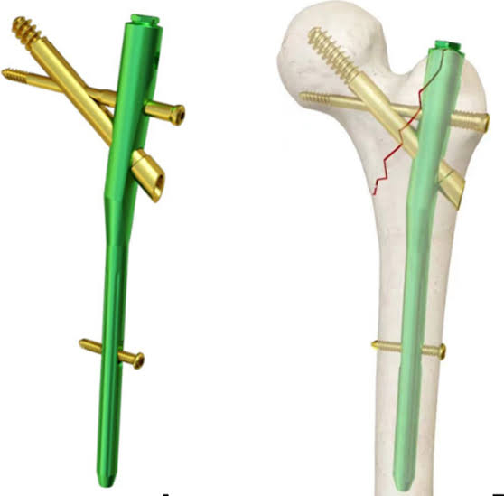
The Proximal Femoral Nail (PFN) is an intramedullary fixation device designed for the treatment of unstable per-, intra-, and subtrochanteric femoral fractures. Unlike extramedullary implants such as the Dynamic Hip Screw (DHS), the PFN is inserted directly into the medullary canal, offering distinct biomechanical advantages through a reduced lever arm and enhanced load-sharing characteristics.
Key Design Features
The PFN incorporates two critical design innovations that differentiate it from earlier intramedullary nails:
- Anti-rotational hip pin: A 6.5 mm hip pin works alongside the main lag screw to reduce the incidence of implant cut-out and provide rotational stability
- Fluted nail tip: The specially shaped nail tip reduces stress concentration, minimizing the risk of low-energy fractures at the distal tip
The distal portion of the nail is available in 10, 11, or 12 mm diameters, while the proximal part measures 17 mm in diameter.
Indications for PFN Nail Surgery
PFN nail fixation is indicated for a range of proximal femoral fractures, particularly those classified as unstable according to the AO/OTA classification system. The procedure is especially suitable for:
- Unstable intertrochanteric fractures (AO/OTA 31-A2 and A3)
- Subtrochanteric fractures
- Reverse obliquity fractures
- Fractures in osteoporotic bone where traditional fixation may fail
The PFN has proven particularly valuable in elderly patients who represent the majority population suffering from these fractures and who benefit most from minimally invasive techniques that preserve fracture hematoma and allow early weight-bearing.
PFN Nail vs. Other Fixation Methods
PFN vs. DHS (Dynamic Hip Screw)
The comparison between PFN and DHS reveals distinct clinical trade-offs. A systematic review published in BMC Musculoskeletal Disorders demonstrated that PFN was associated with shorter hospital stays (average: 7.8 days compared to 12.4 days) and earlier mobilization (7.93 weeks compared to 11.80 weeks to full weight-bearing).
However, PFN was associated with longer fluoroscopy exposure and higher rates of implant cut-out, while DHS showed higher risks for lateral wall fractures, infection, and reoperation. Overall costs proved comparable, with shorter hospital stays offsetting the higher initial implant costs of PFN.
PFN vs. PFNA (Proximal Femoral Nail Antirotation)
The PFNA represents an evolution of the PFN concept, replacing the dual-screw construct with a single helical blade that compacts cancellous bone during insertion. A randomized trial comparing these implants found that PFNA significantly reduces operative time and blood loss compared to conventional PFN.
In osteoporotic patients, PFNA demonstrated superior functional outcomes (PFNA:PFN = 82:75) and fewer implant-related complications, including reduced screw back-out and guidewire breakage. Significant issues in the PFN group included screw back-out, guidewire breakage, and proximal protrusion of the nail tip, while PFNA2 showed more favorable outcomes overall.
PFN vs. Arthroplasty
A meta-analysis of 24 studies involving 2,699 patients found that PFN had significantly shorter surgery duration, less blood loss, and lower mortality compared to arthroplasty for unstable intertrochanteric fractures in the elderly. No significant differences in functional outcomes or complication rates were observed between the two approaches.
Surgical Technique: What to Expect
Preoperative Planning
Successful PFN surgery begins with careful preoperative planning, including:
- Adequate imaging to determine nail diameter, length, and angle
- Assessment of fracture pattern using AO/OTA classification
- Evaluation of bone quality to anticipate fixation challenges
- Consideration of patient anatomy, particularly femoral bow and medullary canal diameter
The Procedure
PFN insertion is accomplished through a minimally invasive technique with a smaller skin incision that preserves the fracture hematoma—an essential element in fracture healing.
Step-by-step overview:
- Patient positioning: Typically supine on a fracture table with the affected limb in traction
- Entry point identification: Through the piriformis fossa or greater trochanter tip
- Guidewire placement: Verified under fluoroscopy before drilling
- Nail insertion: Advanced over the guidewire into the medullary canal
- Proximal locking: Placement of the lag screw and anti-rotation hip pin
- Distal locking: Static or dynamic locking based on fracture stability
The procedure typically takes 1-2 hours to complete under general or spinal anesthesia.
Technical Pearls
Critical technical considerations for optimal outcomes include:
- Proper guidewire placement is verified before drilling to avoid malpositioning
- Screw positioning matters greatly: Implant-related complications were seen mostly in cases of screws placed in peripheral zones and were almost absent in patients with a tip-apex distance (TAD) ≤ 25 mm
- Fracture reduction quality directly impacts outcomes: most patients with acceptable or good reduction did not experience implant-related complications
Clinical Outcomes and Evidence
Fracture Union Rates
The evidence consistently demonstrates that PFN achieves excellent fracture union rates. In a prospective study of 55 patients, fractures united in all patients, with a mean union time of approximately 14.8 weeks for PFN and 14.2 weeks overall.
Most type 3A1 and 3A2 fractures showed radiographic union at 3 months, while type 3A3 fractures typically united by 5 months.
Functional Outcomes
A randomized trial involving 76 patients with unstable intertrochanteric fractures reported excellent Harris Hip Scores in 60.52% of PFN patients and good scores in 31.57%. A separate study noted that PFN patients achieved a mean Harris Hip Score of 71.9 at 3 months and 77.86 at 6 months.
Complication Profile
While PFN is generally well-tolerated, specific complications to monitor include:
- Screw cut-out: More common with improper screw placement or excessive TAD
- Z-effect: A phenomenon unique to dual-screw constructs involving differential screw migration
- Anterior thigh pain: Reported in up to 52% of PFN patients in some series
- Secondary varus: Can occur with inadequate reduction or fixation
One study reported a 27% mortality rate during the first 3 months in elderly patients with unstable trochanteric fractures, though this reflects the frailty of the patient population rather than the implant itself.
Recovery Timeline and Postoperative Care
Immediate Postoperative Period (Days 1-3)
- Hospital monitoring for pain management
- Begin gentle physiotherapy to prevent stiffness
- Early mobilization is typically encouraged
Early Recovery (Weeks 1-6)
- Protected weight-bearing with crutches as advised by the surgeon
- Continued physical therapy focusing on range of motion and strengthening
- Immediate full weight-bearing was permitted in 49 of 55 patients in one prospective study
Full Recovery Timeline
- Fracture union: Typically achieved by 3-6 months
- Full functional recovery: 3-6 months, with continued improvement up to 12 months
- Return to pre-injury function: Approximately 50% of patients achieve similar functional levels to pre-injury status
Postoperative Instructions
Patients should follow these guidelines for optimal recovery:
- Adequate rest with prescribed exercises
- Incision care: Keep the area clean and dry
- Nutritional support: Vitamin and calcium-rich foods to support bone healing
- Pain management: As prescribed by the healthcare provider
- Warning signs to monitor: Severe pain, swelling, redness, or discharge from the incision site
Frequently Asked Questions
What is the success rate of PFN surgery?
PFN surgery demonstrates excellent outcomes with fracture union rates exceeding 90% and good-to-excellent functional outcomes in the majority of patients. The Salvati and Wilson hip function score was 32 out of 40 in 88% of patients at 24-week follow-up in one study.
How long does it take to recover from PFN surgery?
Recovery varies by individual, but patients can expect 3-6 months for full recovery. Early mobilization begins immediately after surgery, with progressive weight-bearing guided by the surgeon.
Is PFN better than DHS?
Both implants have distinct advantages. PFN offers shorter hospital stays, earlier mobilization, and better early functional outcomes, while DHS remains a reliable, cost-effective option for stable fractures with intact lateral walls. The choice depends on fracture type, bone quality, and surgeon expertise.
Can the PFN nail be removed?
Implant removal is typically reserved for specific complications such as infection, nonunion, or significant mechanical failure. Routine removal is not standard practice, as it carries additional surgical risks.
Conclusion
The PFN nail represents a proven, biomechanically sound solution for intertrochanteric femur fractures. Its minimally invasive nature, combined with excellent fracture union rates and favorable functional outcomes, makes it a valuable tool in the orthopedic surgeon’s armamentarium.
While newer designs like the PFNA offer advantages in osteoporotic bone and reduce certain complications, the PFN remains a reliable option—particularly in settings where it is readily available and surgeons are experienced with its use. The key to successful outcomes lies in careful patient selection, precise surgical technique, and attention to detail regarding implant positioning and fracture reduction.
As orthopedic technology continues to evolve, the PFN’s legacy as a foundational intramedullary fixation device remains secure, with ongoing refinements promising even better outcomes for patients with these challenging fractures.